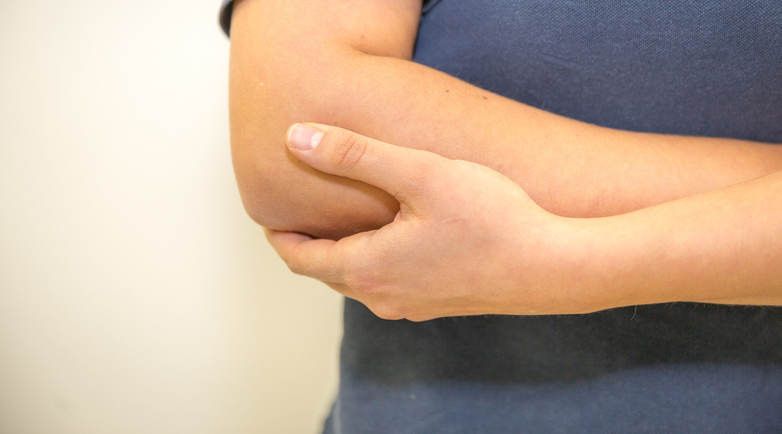
Elbow
The causes of elbow pain are many and varied, including inflammatory reactions due to overuse in everyday life and sports, rheumatic diseases and acute injury. It is important to differentiate the pathology exactly in the history and clinical examination as well as through radiological diagnostics in order to taylor an ideal management plan for it. In addition to conservative treatment, arthroscopic and open surgical procedures are available. The most common pathologies and their therapy are briefly explained below.
Acute injuries of the elbow
Elbow dislocation
The elbow is the second most commonly dislocated human joint, accounting for 20% of dislocations. During dislocation of the elbow a rupture of the capsule-ligamentous structures occurs in all cases (100%). This is followed by cartilage lesions in 30-50% and fractures of the acticulating bones in 20-30%. Rarely vascular (blood vessels, 3-4%) or lasting neurological injuries (3%) accompany this.
After joint reduction, the damage must be evaluated to determine the required treatment. Simple dislocations with single ligament ruptures can usually be treated conservatively.
If more than one primary stabilizing ligament is destroyed, surgical reconstruction of the ligament structures is indicated to prevent chronic instability of the elbow joint. During the operation, the torn ligaments are reattached to the bone in their original position. The anatomical joint function is therefore restored.
Physiotherapy of the elbow begins the day after the operation.
When the swelling of the arm has subsided and the wound has healed, a motion splint with a preset limitation of the range of motion is applied for 6 weeks.
Distal biceps tendon rupture
The tearing of the biceps tendon near the elbow joint at the forearm (radius) can either be caused by an accident or by chronic overload. The loss of strength is obvious when comparing the two sides and the lack of tension on the tendon is proven by the clinical Hook test. Conservative therapy can at best achieve painless movement of the elbow, but the full strength will not be restored. A minimally invasive operation is required to restore this strength for supination (turning the forearm so that the palm of the hand faces forwards) and flexion. As part of the operation, the severed end of the tendon is reattached to its original location on the bone using a suture anchor.
Physiotherapy of the elbow begins directly on the day after the operation.
When the swelling of the arm has subsided and the wound has healed, a motion splint with a preset limitation of the range of motion is applied for 6 weeks.
Cartilage injuries
A cartilage injury to the elbow joint can be caused by direct trauma or shear forces in the context of a dislocation. Frequently there are a combination of injuries to cartilage and bone. Conservative therapy is indicated for small lesions. For larger defects, our primary goal is refixation of the free cartilage-bone fragment. If this is not successful, cartilage therapy using microfracturing, cartilage-bone transplantation (OATS) or transplantation of cultured cartilage cells may be indicated. In most cases, joint function is ensured through these therapeutic methods.
Physiotherapy of the elbow begins directly on the day after the operation.
When the swelling of the arm has subsided and the wound has healed, a motion splint with a preset limitation of the range of motion is applied for 6 weeks.
Plica radialis syndrome
The plica radialis is a soft tissue tongue between the joint capsule, distal humerus and the head of the radius. If the plica is trapped between the bones of the joint, an injury with subsequent pain and swelling may occur. The pain occurs on the outer side of the elbow and may vary during the day, depending on activity. In most cases, this can be treated conservatively by immobilization and medication. If the pain recurs and no improvement can be achieved conservatively, the plica can be removed by arthroscopic surgery.
The rehabilitation period after these operations is short and patients are usually largely symptom-free and able to resume work after 3 weeks.
Chronic diseases and overuse syndromes
Epicondylitis radialis / ulnaris (tennis / golfer’s elbow)
The terms golfer’s elbow or tennis elbow are used to describe pain at the inner or outer protrusion of the elbow. This is caused by an overload of the forearm flexor muscles (golfer’s elbow) or extensor muscles (tennis elbow). The overload can be caused by unilateral activity or by ligament instability of the elbow joint. In the case of chronic overloading, the pain moves from the muscles to the insertion points at the elbow and can lead to structural changes here. The medical history and clinical examination are completed by sonography and magnetic resonance imaging.
Initially conservative treatment (medication/ physiotherapy/ immobilization) is trialled. If the symptoms persist, surgery may be considered. By means of an arthroscopic examination of the elbow joint, structural causes such as instability are uncovered.
Chronic refractory epicondylitis are operated in the technique described by Nirschel. This leads to a complete reconstruction and stabilization of the muscle attachments. This is an important prerequisite for a strong and well-functioning elbow joint.
Physiotherapy to the elbow begins directly on the day after the operation.
When the swelling of the arm has subsided and the wound has healed, a motion splint with a preset limitation of the range of motion is applied for 6 weeks.
Elbow instability (radial/ulnar)
If a capsular/ligamentous instability of the elbow joint is determined by clinical examination, the elbow can be stabilized through specific physiotherapy. If an instability remains despite conservative treatment, diagnostic arthroscopy is indicated. By means of this the insufficient ligament can be identified and treated by ligamentoplasty.
Physiotherapy of the elbow starts directly on the day after the operation.
When the swelling of the arm has subsided and the wound has healed, a motion splint with a preset limitation of the range of motion is applied for 6 weeks.
Loose bodies (LB)
Loose bodies in the elbow are often a result of accidents or primary joint disorders (e.g. osteochondrosis dissecans).
The LB can lead to crepitation, locking and pain. In addition to the clinical examination, X-rays and, if necessary, computer or magnetic resonance tomography are performed to make the diagnosis.
In almost all cases, the removal of the LB can be performed arthroscopically.
Physiotherapy of the elbow begins directly on the day after the operation.
Joint stiffness of the elbow (arthrofibrosis)
Stiffness of the elbow is usually post-traumatic or arthritic. The extent of disability in everyday life caused by the restricted movement is subjective and individual to the demands of the patient. Even 20° loss of flexion can severely inhibit daily activities. With the help of conservative therapy, with physiotherapy and specific splints, a large proportion of functional limitations can be eliminated. However, if sufficient mobility cannot be achieved by exercising the joint, adhesions and bony spurs can be removed during arthroscopic or open surgery to allow further mobilization .
Physiotherapy of the elbow starts directly on the day of the operation twice daily and is required intensively for at least 6 weeks to ensure a good postoperative result.
Arthritis of the elbow joint
Elbow arthritis is characterized by pain on movement and at rest, as well as an increasing limitation in the range of movement.
Arthritis of the elbow can be treated conservatively to a certain extent. Depending on the location, there are a number of joint-preserving interventions to eliminate the symptoms of arthritis or to halt its progression. Arthroplasty (implantation of an artificial joint) is the final step after a series of conservative and surgical joint-preserving measures. The entire joint, or only the most diseased areas of a joint may be replaced by an artificial surface. In all cases, the aim is to restore good pain-freeelbow function.
Physiotherapy of the elbow begins directly on the day after the operation.
When the swelling of the arm has subsided and the wound has healed, a motion splint with a preset limitation of the range of motion is applied for 6 weeks.


 Contact for Patients
 Phone +49-(0)89 4140-7840 /-7830
